Ultrasound uses sound waves to view organs and soft tissues. The probe is placed on the skin and images appear immediately, so the doctor can observe structures during the examination itself. No radiation is involved, the study is commonly used in children and during pregnancy.
It is requested for abdominal or pelvic symptoms, swelling, thyroid and joint problems, and routine pregnancy checks. The same method is used to observe fetal growth and position. A Doppler study is added when the blood flow in vessels or the heart needs assessment. Ultrasound may also guide needle placement during certain procedures.
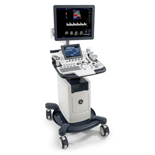
The examination is performed on 2D/3D systems with a colour Doppler facility. A radiologist conducts the scan, while a cardiac echo is performed by a cardiologist.
Gel is applied over the area, and the probe is moved slowly to obtain different views. The test is usually completed within a short time and does not cause pain.
Need of USG :
To diagnose an individual who has symptoms in certain tissues or organs. These include the heart, kidney and also the female reproductive system.
To conduct a biopsy, an ultrasound can act as a guide so as to confirm where the sample is to be taken from.
To evaluate abdominal pain and diagnose abdominal symptoms in children, such as injuries or appendicitis.
To conduct an echocardiogram to diagnose problems with the heart and to perform an echocardiogram of the fetus to check for heart defects
To gain additional information during pregnancy, such as the age of the developing fetus, the baby’s position and overall health. A prenatal ultrasound can also give the baby’s sex.
We use USG imaging to diagnose multiple diseases, lesions, causes of pain, to evaluate blood flow.
USG is important modality to assess fetal growth through various parameters as well as to assess fetal blood flow.
USG is also used for guiding tool for multiple interventional procedures.
Doctor puts probe on your body area which needs to be scanned and moves it in order to get internal organ’s images from multiple angles (sections). USG probe sends high frequency pulse and captures it’s echo, then USG machine process this data and creates images on machine’s screen.