Comprehensive X-ray and CT scan services supporting accurate diagnosis across clinical needs.
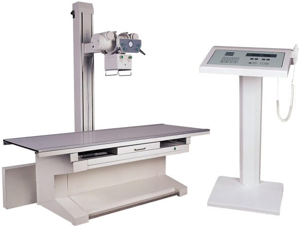
Diagnostic Precision with Advanced X-Ray Solutions
An X-ray is usually the first scan advised for injury, chest complaints, or joint pain. The exposure itself lasts only a moment, and the image is available for immediate viewing.
Doctors use it to check for fractures, lung infection, fluid in the chest, and certain abdominal findings. In many cases, a single image is sufficient. A repeat study is done only if comparison is required during recovery or follow-up.
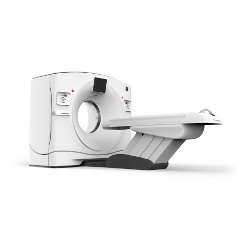
Computed Tomography (CT Scan)
A CT scan is requested when the area cannot be assessed adequately on X-ray or when deeper structures must be seen. The scanner records thin sections of the body, which are reviewed together rather than as a single picture.
It is commonly performed in head injury, suspected stroke, severe abdominal pain, and trauma. The study also helps in monitoring known diseases over time.
Images are examined by a radiologist with the clinical notes before the report is issued to the treating doctor.