Viral ( HHV6) encephalitis - A case report
Section:
Neuroradiology
Case Type:
Clinical Cases
Authors:
Dr. Parth Patel, Dr. Madhuri Ghate
Email: parthpatel1302182@gmail.com, drmadhuri.ghate@krsnadiagnostics.com
Patient:
40 Years, female
Clinical History:
- A 40 year-old female presented with
- H/O SUDDEN ONSET UNCONSCIOUSNESS
- H/O FEVER /VOMITING
- H/O GENERAL BODY PAIN/WEAKNESS
- NO H/O TRAUMA /FALL
Imaging Findings:
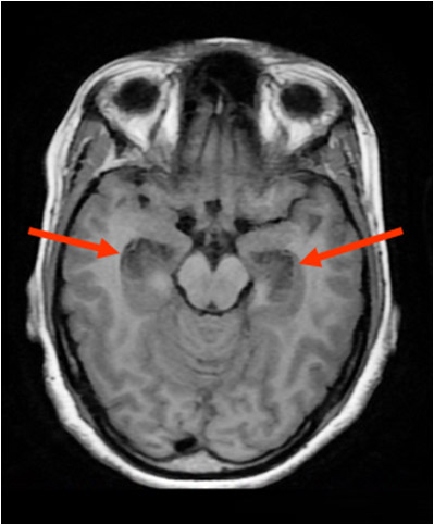
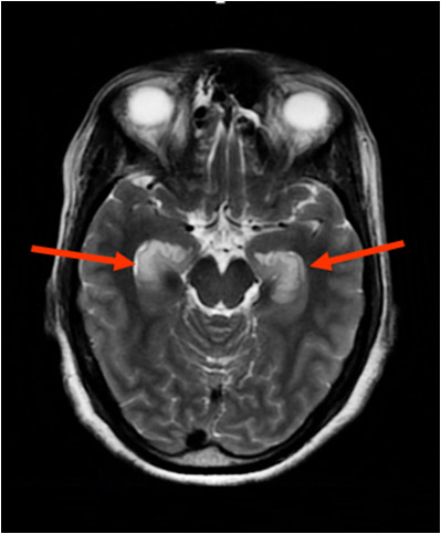
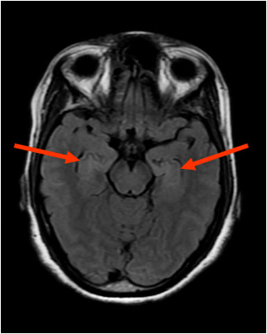
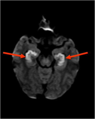
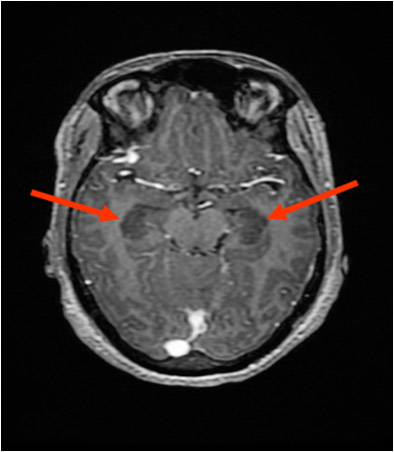
Plain and contrast MRI Brain was advised which showed Bilateral Symmetrical areas of cortical thickening with edema in bilateral medial temporal lobes, limbic cortex and hippocampus. It appears as hypo-intense signal on T1 WI , hyper-intense signal on T2 and T2 FLAIR images. It shows and restricted diffusion with corresponding low ADC values. No obvious enhancement noted post contrast. (Fig. 1, 2, 3, 4 and 5- red arrow).
FIGURE 2
AXIAL MRI BRAIN T2WI
Axial T2WI MRI shows hyper intense signal in bilateral Medial Temporal lobes, Limbic cortex and Hippocampus ( Red arrow)
FIGURE 3
AXIAL MRI BRAIN T2 FLAIR IMAGE
Axial T2 FLAIR MRI shows hyper intense signal in bilateral medial Temporal lobes, Limbic cortex and Hippocampus (red arrow)
FIGURE 4
AXIAL MRI BRAIN DIFFUSION WEIGHTED IMAGE
DWI MRI image shows restricted diffusion in bilateral medial Temporal lobes, Limbic cortex and Hippocampus (red arrow)
FIGURE 5
AXIAL MRI BRAIN APPARENT DIFFUSION COEFFICIENT IMAGE
ADC MRI image shows low ADC value noted in bilateral medial Temporal lobes, Limbic cortex and Hippocampus (pointed by red arrow)
FIGURE 6
AXIAL MRI BRAIN T1WI WITH CONTRAST
Axial MRI brain T1WI with contrast shows no Enhancement (pointed by red arrow)
