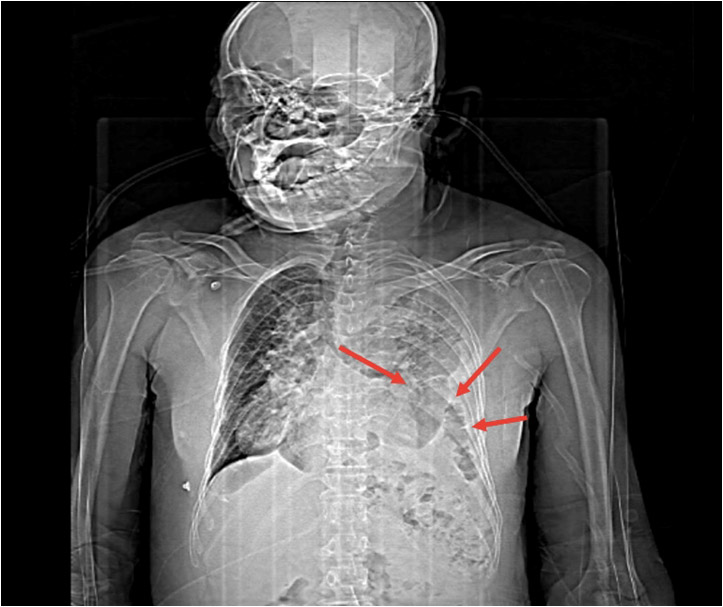
Left Diaphragmatic Hernia - A Case Report
Section:
CHEST IMAGING
Case Type:
CLINICAL CASES
Authors:
DR SAGAR ANTALA, DR MADHURI GHATE
EMAIL- sagarsahil677@gmail.com , drmadhuri.ghate@krsnadiagnostics.com
Patient:
60 YEARS, MALE
Clinical History:
A 60 years old male presented with H/O blunt trauma of abdomen and thorax associated with breathlessness, tachypnea and coughing.
Imaging Findings:
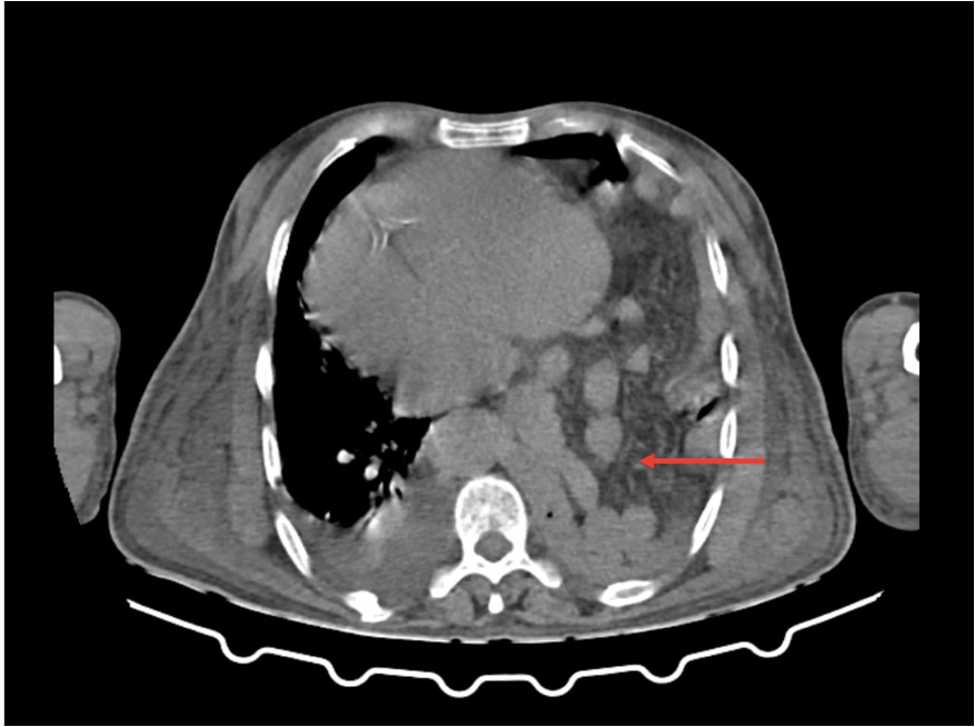
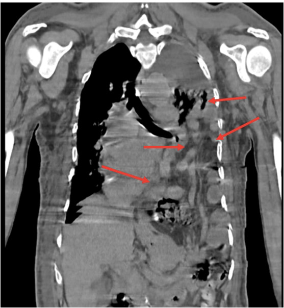
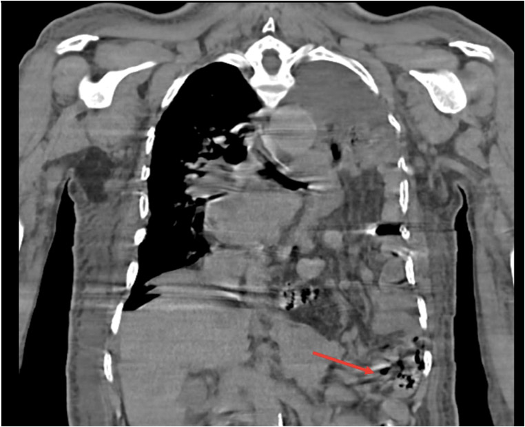
- Plain HRCT Thorax was advised which showed A defect in posterolateral left hemidiaphragm with intrathoracic herniation of splenic flexure of colon, small bowel and vast amounts of the mesentery associated with cardiac, tracheal and mediastinal shift towards right side. (Fig. 1, 2, 3 and 4 - red arrow)
- Moderate to severe left pleural effusion is noted with underlying atelectatic changes and consolidation and reduced lung volume.
- Mild to moderate right sided pleural effusion with underlying atelectatic changes also noted.
- No mediastinal haematoma or pneumomediastinum is noted and the mediastinal fat planes are well maintained and appear normal.
Differential Diagnosis:
- LEFT DIAPHRAGMETIC HERNIA
Final Diagnosis:
LEFT DIAPHRAGMETIC HERNIA (TRAUMATIC)
FIGURE 2
Fig 2 -AXIAL SECTION OF PLAIN CT-THORAX IN MEDIASTINAL WINDOW shows herniation of small bowel and mesentery in left hemithorax.(red arrow)
FIGURE 3
Fig 3 CORONAL SECTION OF PLAIN CT-THORAX IN MEDIASTINAL WINDOW shows intrathoracic herniation of splenic flexure of colon, small bowel and vast amounts of the mesentery. (red arrows)
FIGURE 4
Fig 4-CORONAL SECTION OF PLAIN CT-THORAX IN MEDIASTINAL WINDOW shows defect in left hemidiaphragm. (red arrows)


