Neuroimaging
Section:
Spine imaging
Authors:
Dr. Akash Patel , Dr.Madhuri Ghate Krsnaa Diagnostics PVT LTD; Pawana Nagar Housing Society 411033 Chinchwad, India; Email:drmadhuri.ghate@krsnadiagnostics.com
Clinical History:
35 years old male with seizures
Imaging Findings:
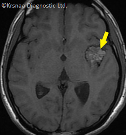
- Fig. 1 – Axial T1WI of brain shows well defined hyperintense rounded lesion with hypointense surrounding rim in left temporal lobe (yellow arrow)
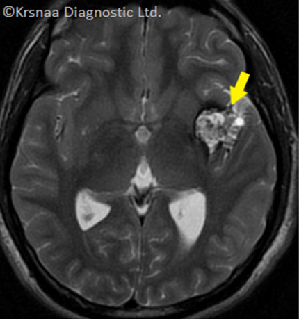
- Fig. 2 – Axial T2WI of brain shows well defined hyperintense rounded lesion with hypointense surrounding rim in left temporal lobe giving typical popcorn appearance (yellow arrow)
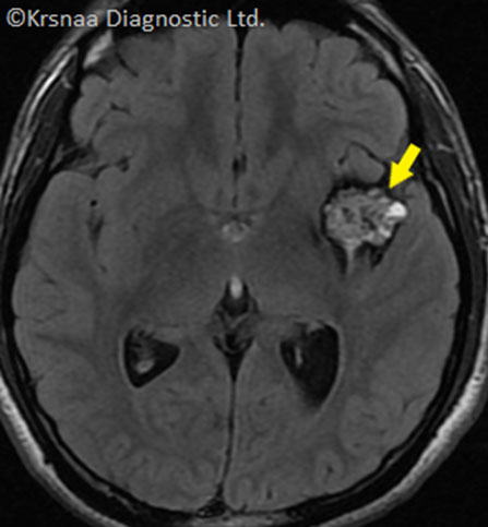
- Fig. 3 - Axial T2 FLAIR image of brain shows well defined hyperintense rounded lesion with hypointense surrounding rim (yellow arrow)
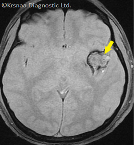
- Fig. 4 - Axial GRE image of brain shows peripheral blooming of lesion.
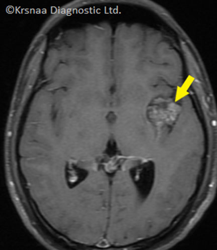
- Fig. 5 - Axial T1 Post contrast image of brain shows mild faint enhancement of the lesion (yellow arrow)
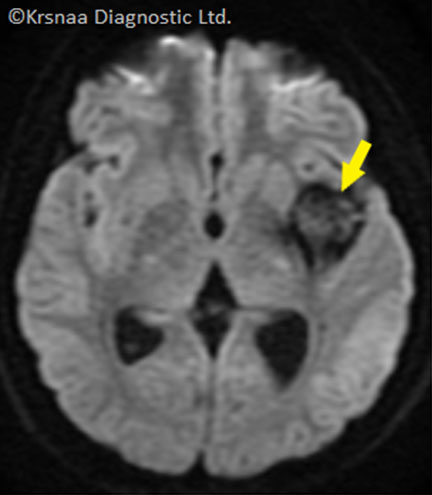
- Fig. 6 - Axial DWI of brain shows no restricted diffusion
Differential Diagnosis:
- Cavernous angioma
- Haemorrhagic metastasis
- Ependymoma
- Glioblastoma
- Toxoplasmosis
- Arteriovenous malformation
Final Diagnosis:
Cavernous angioma - Zambraski Type II
