Pagets Disease
Section:
Head imaging
Spine imaging
Authors:
Dr.Miral Patel, Dr.Madhuri ghate Krsna Diagnostics PVT LTD; Pawana Nagar Housing Society 411033 Chinchwad, India; Email:drmiralpatel1995@gmail.com, drmadhuri.ghate@krsnadiagnostics.com
Clinical History:
60 yrs old male with headache and giddiness.
Imaging Findings:
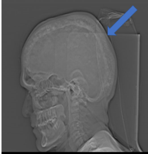
- Scout image (fig:1)of CT brain shows widening of diploic space of skull bones(blue arrow) .
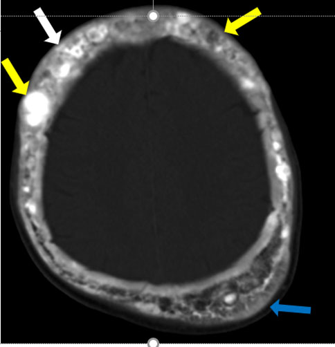
- Axial section of CT brain, bone window (fig:2), shoes multiple lytic and sclerotic lesions(yellow arrows) in skull with widening of diploic spaces(blue arrow) giving cotton wool appearance(white arrow).
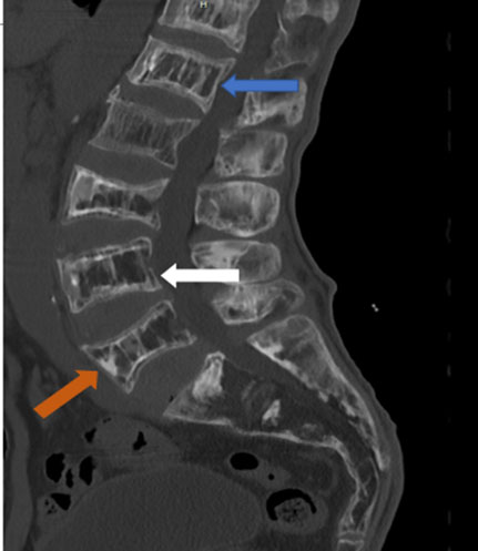
- Sagittal section of CT Spine(fig :3) shows cortical thickening and sclerosis encasing the vertebral margins(blue arrow) with coarsening of trabaculae in vertebral bodies(brown arrow).[Picture frame sign; white arrow]
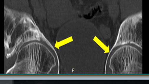
- Visualised portion of pelvis bone in coronal section of CT (fig 4)show acetabular protrusion(yellow arrows).
Differential Diagnosis:
- For skull lesions: fibrous dysplasia pagets disease
- For spinal lesions : vertebral haemangioma
Final Diagnosis:
Pagets disease
FIGURE 2
Fig:2 Axial section of CT brain ( bone window)shoes multiple lytic and sclerotic lesions(yellow arrows) in skull with widening of diploic spaces(blue arrow) giving cotton wool appearance(white arrow) ©Krsnaa Diagnostics Ltd.
FIGURE 3
Fig 3 : Sagittal section of CT Spine shows cortical thickening and sclerosis encasing the vertebral margins(blue arrow) with coarsening of trabaculae in vertebral bodies(brown arrow).[Picture frame sign; white arrow] ©Krsnaa Diagnostics Ltd.
FIGURE 4
Fig:4 Visualised portion of pelvis bone in coronal section of CT show acetabular protrusion(yellow arrows) ©Krsnaa Diagnostics Ltd.


