Role of routine and limited MRI imaging in diagnosis and screening of femoral head AVN

Congress:

ECR 2018

Poster Number:

C-096

Type:

Educational Exhibit

Keywords:

Musculoskeletal system, MR, Education, Education and training

Authors

Dr M.S. Ghate, Dr R.R. Kotkar, Dr G.H.Kadam
Krsna Diagnostics PVT LTD; Pawana Nagar Housing Society 411033 Chinchwad, India; Email:madhuri.ghate01@gmail.com

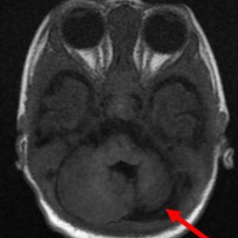
FIGURE 1

Axial MRI BRAIN T1W image
Unilateral Sidebar

Axial T1W MRI image shows left cerebellar hypoplasia (arrow).

Learning objectives

Explain the pathophysiology of AVN. Classifying AVN based on MR imaging findings. Use of limited MRI imaging in screening as well as in early detection of AVN.

BACKGROUND

Avascular necrosis(AVN) or Osteonecrosis of the femoral head occurs due to compromise in vascular supply. Early diagnosis is of utmost importance as delay in diagnosis increases cost of treatment and morbidity.MRI is the sensitive imaging tool for detection of AVN. Our review will focus

Findings and procedure details

PATHOPHYSIOLOGY OF AVN AND ITS IMAGING CORRELATION : Osteonecrosis, also known as avascular necrosis or AVN, is characterized by a stereotypical pattern of cell death due to vascular compromise and a complex repair process of bone resorption and formation. (1) .It is not the necrosis itself but rather the resorptive component of the repair process that results in loss of structural integrity and subchondral fracture. Decreased femoral head blood flow can occur through three mechanisms: vascular interruption by fractures or dislocation, intravascular occlusion from thrombi...

CONCLUSION:

MRI is a sensitive imaging tool for early detection and follow up of AVN.Limited MRI protocol using coronal T1W sequence can be used for screening and early detection which is cost effective and saves time.

Personal information

Contact details: Dr Madhuri Ghate Consultant Radiologist KRSNA DIAGNOSTICS PVT LTD CHINCHWAD , PUNE Email: madhuri.ghate01@gmail.com Dr Rashmi Kotkar Consultant Radiologist KRSNA DIAGNOSTICS PVT LTD CHINCHWAD , PUNE Email : drrashmikotkar@gmail.com INSTITUTION Krsnaa Diagnostics Pvt. LtdPawana Nagar Housing Society, Chinchwad, Pawana Nagar Housing Society, Chinchwad, Pimpri-Chinchwad, Maharashtra 411033Phone:20 27350025/26/27E-mail:Info@krsnadignostics.com

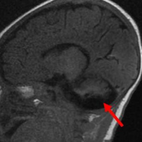
FIGURE 2

Sagittal MRI BRAIN T1W image

Unilateral Sidebar

T1W sagittal MRI image shows left cerebellar hypoplasia.

FIGURE 3

MRI BRAIN Coronal T2W image
Unilateral Sidebar

T2W coronal MRI image shows left cerebellar hypoplasia (arrow).

FIGURE 4

MRI BRAIN Coronal 3D SPGR image

Unilateral Sidebar

3D SPGR coronal MRI image shows left cerebellar hypoplasia (arrow).

TOP
FeedBack
close slider