Spine imaging
Section:
Spine imaging
Authors:
Dr. Akash Patel , Dr.Madhuri Ghate Krsnaa Diagnostics Ltd. ; Pawana Nagar Housing Society 411033 Chinchwad, India; Email : drakash294@gmail.com, drmadhuri.ghate@krsnadiagnostics.com
Clinical History:
27 yrs old male with low backache and leg weakness since 3 month.
Imaging Findings:
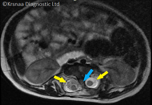
- Fig. 1 Axial section of T2-W MRI of spine shows bony bar (Blue arrow) splitting the spinal cord into two part & creating two distinct spinal canals (Yellow arrow)
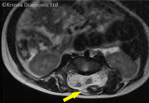
- Fig. 2 Axial T2-W MRI of the spine, shows midline bony defect at the posterior arch - spina bifida. ( Yellow arrow )
Differential Diagnosis:
- Diastometamyelia
Final Diagnosis:
Diastometamyelia Type 1


