Unilateral cerebellar hypoplasia – A case report
Section
Neuroradiology
Case Type
Clinical Cases
Authors
Madhuri Ghate, Rashmi Kotkar, Amol Gulhane
Krsna Diagnostics PVT LTD; Pawana Nagar Housing Society 411033 Chinchwad, India; Email:madhuri.ghate01@gmail.com
Patient
2 months, male
Categories
Area of InterestNeuroradiology brain ; Imaging Technique MR
Procedure Education ; Special Focus Seizure disorders ;
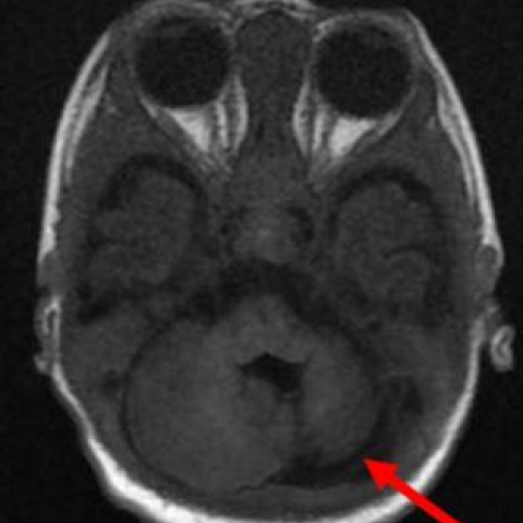
FIGURE 1
Axial MRI BRAIN T1W image

Axial T1W MRI image shows left cerebellar hypoplasia (arrow).
Clinical History
A 2-month-old male child presented with seizures.
Imaging Findings
Plain MRI brain was advised which showed that the left cerebellar hemisphere was hypoplastic and was smaller in size compared to the right side (Fig. 1, 2, 3 and 4 – red arrow). The grey and white matter of the cerebellum was normal and did not show any signal changes. The fourth ventricle was normal. The cisterna magna was normal. The vertebral and basilar arteries showed normal flow voids.
Discussion
Cerebellar malformations are classified into two categories: hypoplasias and dysplasias [1]. Hypoplasias: Those associated with hypoplasia have reduced cerebellar volume. Dysplasias: they have abnormal foliation, and abnormal architecture of the cerebellar white matter. Cerebellar hypoplasias are classified as focal or generalised. Focal being further classified into isolated vermian hypoplasia and hypoplasia of one cerebellar hemisphere. Patients with unilateral cerebellar hypoplasia can be asymptomatic or can present with cerebellar symptoms, headache or rarely with seizure [2]. Aetiopathogenesis is postulated as intrauterine or perinatal vascular insult. Hence hypoplasia or aplasia of the cerebellar or vertebral arteries can be seen in these cases [3, 4]. Intrauterine insult can be a prenatal haemorrhagic lesion, as cerebellum is vulnerable to bleeding during fetal development, which can be diagnosed on prenatal fetal MRI [5]. Intrauterine insult may be due to congenital cytomeglovirus infection, maternal diabetes mellitus, maternal alcohol addiction and placental thrombotic vasculopathy and infarctions. Other factors may play a role in aetiopathogenesis like genetic mutations with somatic mosaicism. Cerebellar hypoplasia can have syndromic association with PHASES syndrome (posterior fossa anomalies, haemangioma, arterial anomalies, cardiac abnormalities/aortic coarctation, eye abnormalities) syndrome [6] and Mobius syndrome. In present case, there was no other structural abnormality found except for isolated unilateral cerebellar hypoplasia.
Differential Diagnosis List
Unilateral cerebellar hypoplasia.
Global cerebellar hypoplasia
Cerebellar hypoplasia with mainly vermis involvement
Final Diagnosis
Unilateral cerebellar hypoplasia.
FIGURE 2
Sagittal MRI BRAIN T1W image

T1W sagittal MRI image shows left cerebellar hypoplasia.
FIGURE 3
MRI BRAIN Coronal T2W image
T2W coronal MRI image shows left cerebellar hypoplasia (arrow).
FIGURE 4
MRI BRAIN Coronal 3D SPGR image