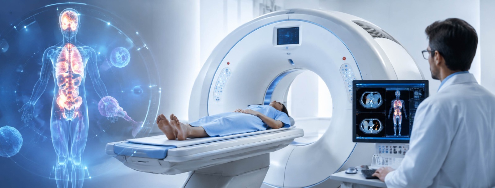
Oncologic workflows move fast. A PET-CT scan maps tumours, tracks therapy progress, and flags recurrence early. Knowing the baseline steps cuts hospital anxiety. The machine blends two distinct imaging styles. A CT scanner maps anatomy via X-rays. Simultaneously, an intravenous radiotracer, usually fluorodeoxyglucose, enters the bloodstream. Malignant cells burn glucose rapidly, pulling the tracer straight into active lesions.
Oncologists rely on this dual data to check nodal spread. It measures mid-cycle shrinkage, catching relapse long before physical symptoms surface. Protocols vary. Standard tracers fit routine checks, but specialised exams, like PSMA scans for prostate issues or DOTATATE scans for neuroendocrine tumours,
require short-lived isotopes brewed fresh that morning. Whole-body checks differ from targeted brain or heart sequences.
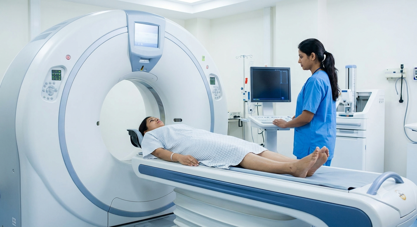
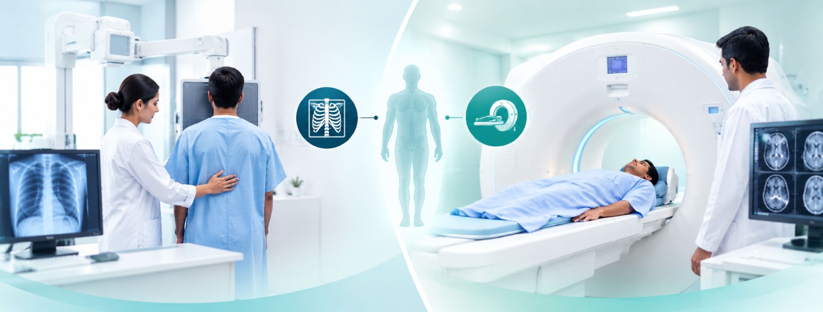
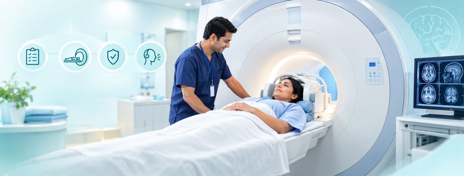
Prep dictates success. Fasting four to six hours drops blood sugar so the tracer hits cancer targets instead of circulating glucose. Diabetics get custom medication rules. Next comes the wait. After the IV line goes in, patients sit quietly in a low-stim room. Moving makes muscles soak up the tracer instead of the tumour. Once that window closes, you lie flat on a motorised table. The gantry hums. Absolute stillness is mandatory for thirty minutes.
The wait for results wears on patients. Anxiety spikes between the scan and the follow-up chat, making a patient advocate valuable during oncology consults to break down complex metrics like standardised uptake values. India’s top hospitals match global imaging standards, but true success relies on human collaboration. When your nuclear team, medical oncologist, and surgeons talk openly, raw scan data turns into a clear, personalised recovery plan.
Clinical teams review these parameters constantly to adjust care paths. Every phase from initial tracer uptake to final multidisciplinary evaluation ensures that diagnostic insights translate smoothly into actionable therapeutic decisions for the patient.