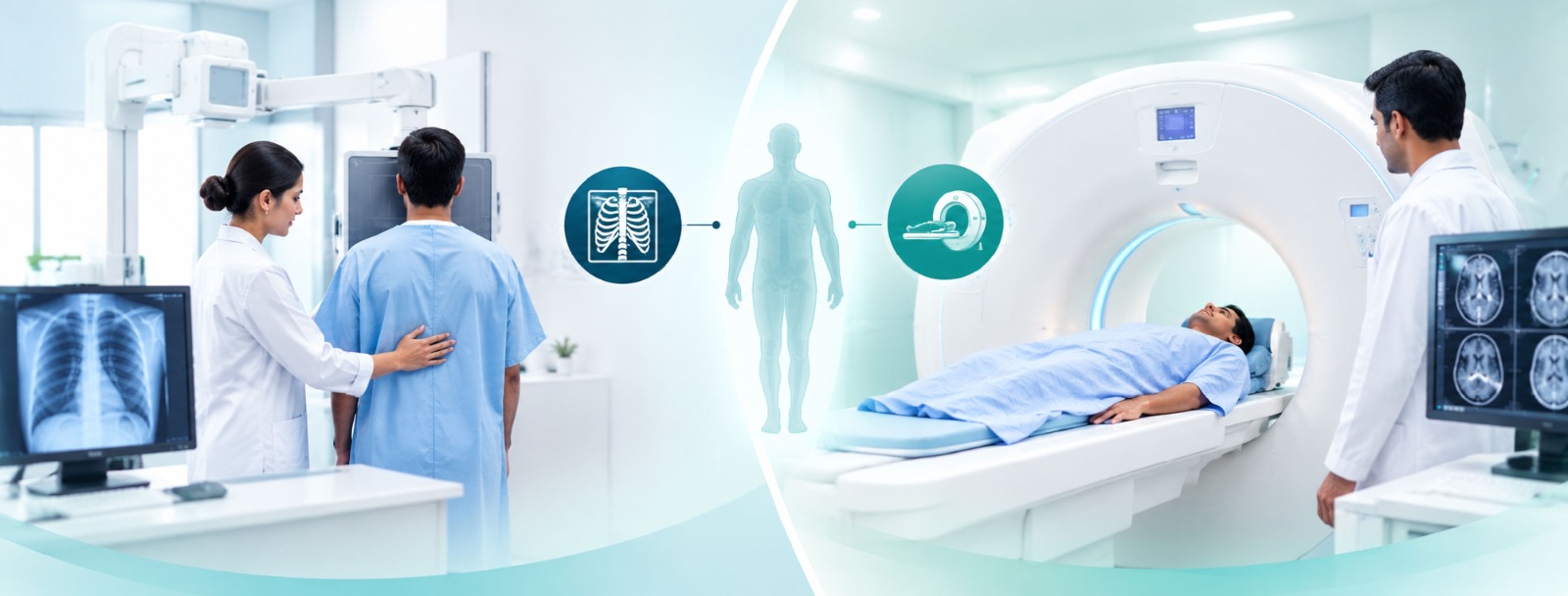
Have you ever walked into a hospital radiology wing? It is freezing, smells entirely too clean, and they just shove you toward these giant, intimidating metal tubes without giving you the slightest clue why. They point at some high-tech monstrosity and tell you to wait. Total nightmare. Hospitals are completely packed with this heavy gear, and every single machine, whether we are talking X-rays, ultrasounds, CTs, MRIs, or PET scans, uses totally different physics to figure out what is wrong with your body. It is actually wild if you take a step back and think about how they pull it off.
Let us start with X-rays. Old school. Classic. A technician slaps a digital plate behind your back, zaps a quick burst of radiation right through your torso, and bam, you have a basic shadowgram. Dense bone stops the beam cold, so it looks blindingly white on the screen. Soft tissue? The radiation slides right through, leaving it dark. People always freak out about radiation risks, but honestly, a standard flight across the country exposes you to way more cosmic rays than a normal chest X-ray. It is cheap, blazing fast, and killer for catching broken bones. But if you are looking for a soft tissue tear? X-rays are completely, utterly blind.
Then you have ultrasounds. No radiation here at all, which is a huge relief. They just use high-frequency sound waves. The technician smears that freezing cold gel directly onto your stomach or chest, slides a plastic probe around, and the machine listens carefully to the echoes bouncing off your insides to draw a live, moving video. Everyone knows it from pregnancy checkups, but doctors use it constantly for heart valves, blood flow, and guiding biopsy needles. The downside is pretty brutal, though: sound waves hit bone or gas-filled pockets and just stop dead. You cannot use this to look deep inside a skull or peer through a lung. Total physical roadblock.
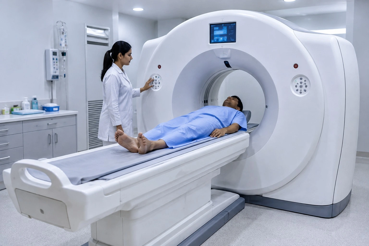
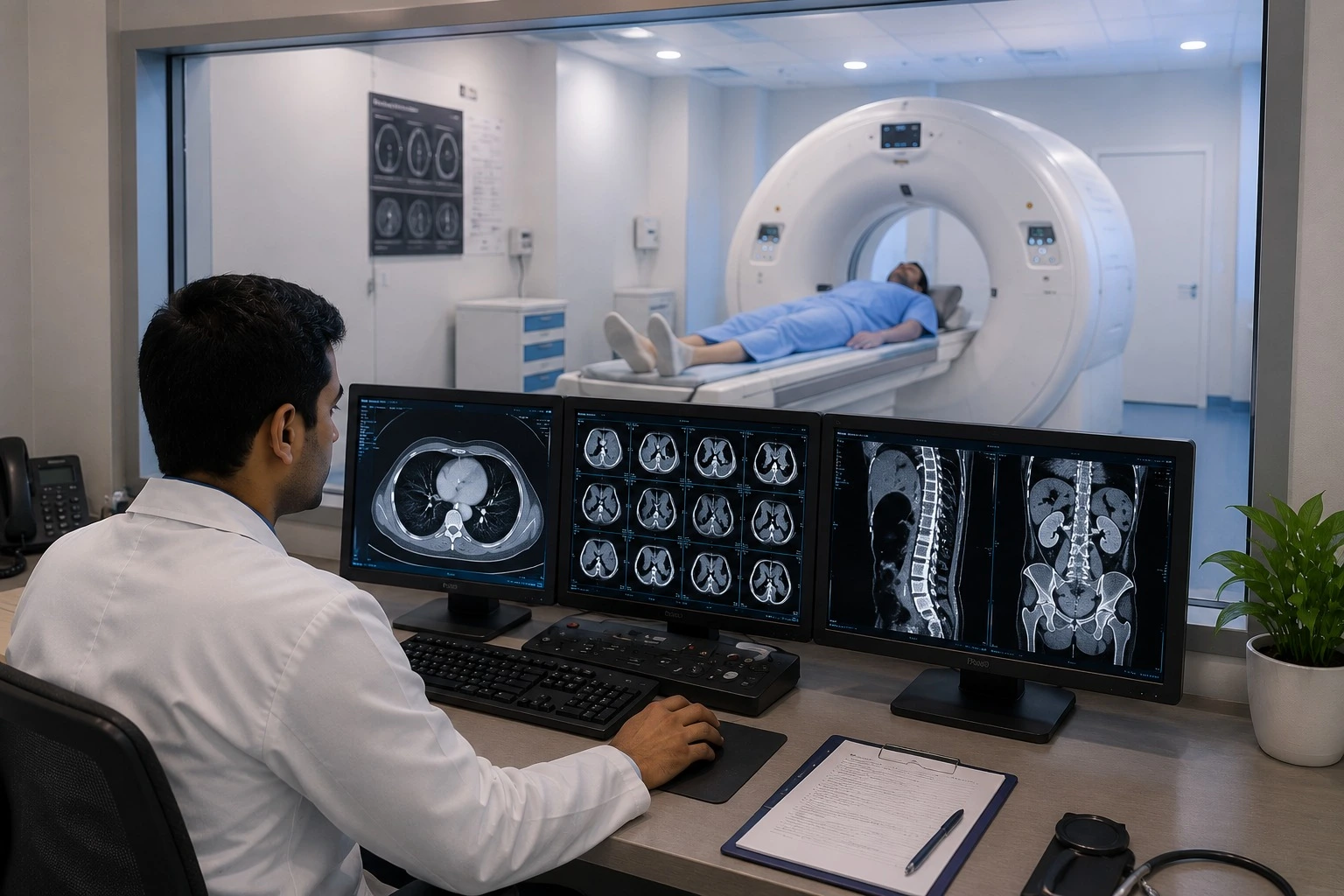
CT scans are basically X-rays on absolute steroids. The scanning ring spins around your body in a hyper-fast circle, grabbing dozens of different slice angles in mere seconds. A powerful computer stitches all those individual angles together into a detailed 3D model of your insides. Emergency room doctors live and breathe by CT scans because they take seconds and can instantly flag a dangerous brain bleed or a deadly blood clot in the chest. The dark side? The radiation dose is substantially heavier than a normal X-ray, so physicians try to avoid ordering them unless things are genuinely an acute emergency.
MRIs are where medical imaging gets genuinely weird. Zero radiation. Instead, they swap it for insanely powerful superconducting magnets and radio pings. The machine forces every single hydrogen atom in your body into a strict, uniform line, then knocks them out of place just to listen to the magnetic snapback. The level of detail on cartilage, ligaments, and brain tissue is completely unmatched;
it is the gold standard for neurology and orthopaedics. But the experience? Absolutely miserable. You are trapped inside a loud, claustrophobic tube for forty minutes listening to hammering noises, and if you have unverified metal or a pacemaker in your body, you are straight-up barred from entering the room.
PET scans mix metabolic tracking with a traditional CT setup. Nurses pump a tiny dose of radioactive sugar into your bloodstream, and the camera hunts down where the glucose pools up. Why? Because runaway cancer cells and gnarly infections burn through sugar like absolute wildfire. It is not great for showing fine anatomical shapes on its own, but it catches chemical shifts and cellular chaos long before a tumour physically wrecks an organ.
At the end of the day, picking the right scan always comes down to what the doctor is hunting for. Broken bones and lung checks start with simple X-rays. Soft tissue motion goes straight to ultrasound. Acute trauma demands a CT. Complex joints and brains need an MRI. Cancer staging calls for a PET scan. It is not magic, even if the equipment looks like it walked right off a spaceship.